By A Mystery Man Writer

Imaging echogenic breast masses. - Document - Gale OneFile: Health
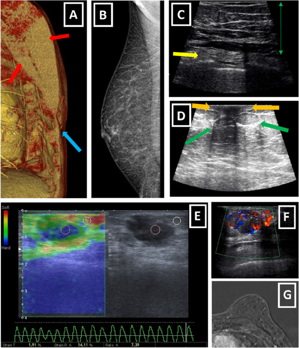
Imaging findings and classification of the common and uncommon

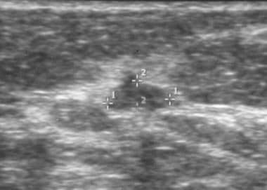
e Ultrasound images of mass. (A) Antiradial ultrasound image of

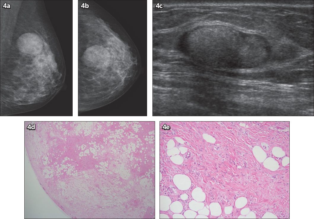
The ultrasound and mammogram of a 34 year-old woman 2 months after
Breast Cancer Ultrasonography: Practice Essentials, Role of

a. Case 1. Right breast USG showing well-defined, hypoechoic mass

Breast – Something About Radiology – Just For Sharing

PDF) Tumoral pseudoangiomatous stromal hyperplasia: Radiological
Four cases of echogenic breast lesions: a case series and review
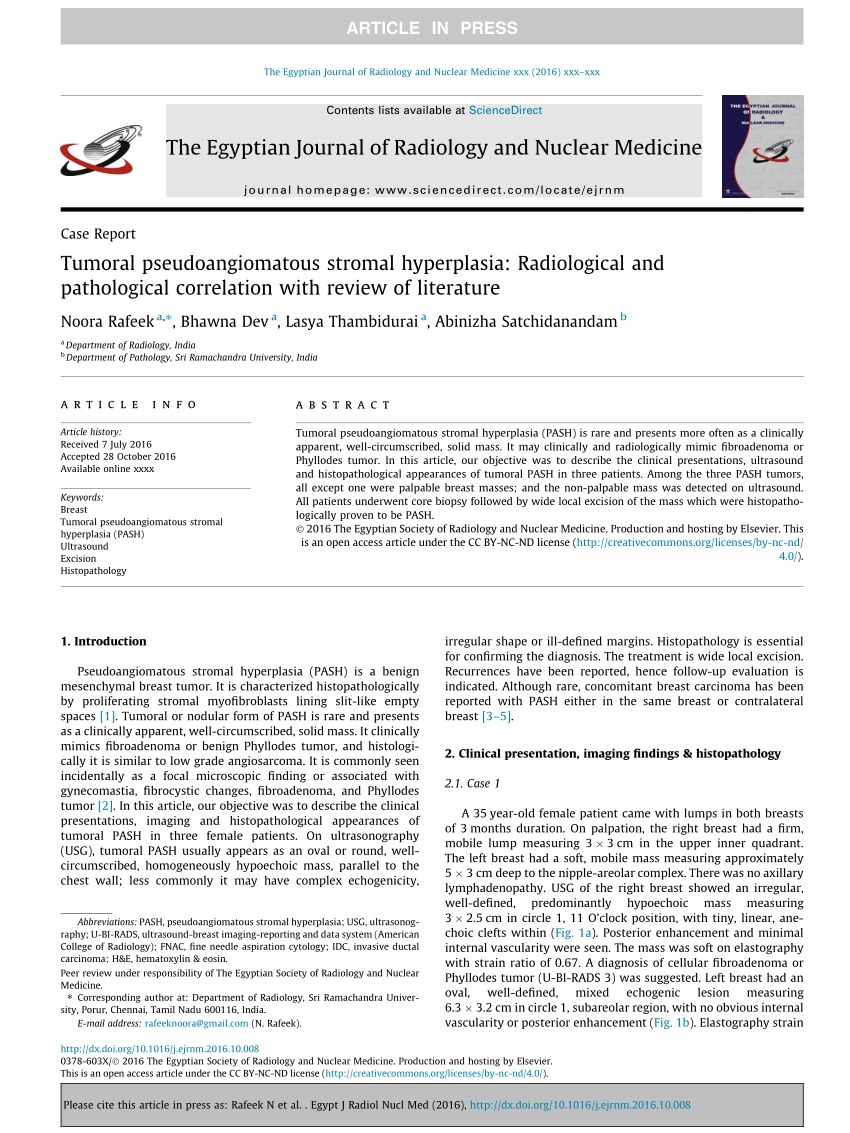
PDF) Tumoral pseudoangiomatous stromal hyperplasia: Radiological

Breast mass, Radiology Reference Article
The Breast SpringerLink